Patients who have experienced a traumatic brain injury may have delayed pupil reactivity. Color vision may also be an issue for them in certain situations.
The basic idea about brain injury and pupil reactivity or pupillary size measurement
It is triggered when light contacts the retina and stimulates cranial nerves 3, 6, 9, 10, and 11 to send instructions to the brain. This is an innate pupillary light reflex. Patients who have had significant brain damage may have difficulty seeing these objects due to impairment to the optic nerve area or other surrounding sites.
Doctors may assess the pupillary light response by shining an intense light into the patient’s eye for a short time during the evaluation of pupillary reaction. Patients who have been injured may not be able to close their eyes while still seeing.
Patients’ linguistic abilities and motor skills, such as balance and coordination, are examined to determine how well their brains are working after traumatic brain injury. An example of a standardized test is the mini-mental state examination (MMSE), which asks questions on the quantity of information being processed at any given time (MMSE).
It is necessary to test patients’ cognitive abilities using the MMSE to help doctors determine how much function has been lost due to the damage. Doctors may use the results of this pupillary response in traumatic brain injury as a starting point for further studies of brain function, and they may also consult with patients. When this information is accessible, it is also possible to provide medications such as gabapentin, which may help with speech and brain function without risk.
Other Tests
Other traumatic brain injury diagnostic tests include: The Glasgow Coma Scale is used to determine how much function has been lost in a person suffering from a traumatic brain injury (GCS). It includes an eye-opening, verbal response, and muscle response, among other things. Patients are considered to be in a coma if none of the three types of responses come back; patients are said to be in a “unilateral coma” if only one category responds; patients are said to be in a “bilateral” coma if two or more types reply to the questionnaire.
A clinician’s ability to assess the severity of brain injury and determine which areas of the brain are still functioning is enhanced by using the GCS. Additional tests, such as the Mini-Mental State Examination (MMSE), may be performed in combination, although they are not required for this assessment.
It is necessary to assess patients’ ability to follow a penlight as it moves across their face, as well as their ability to react to different lights or shapes that are flashed in front of their eyes during a pupillary examination. The behavior of a traumatic brain injury patient to a visual acuity test may involve looking at different letter or form sizes from close ranges in response to the test. This procedure may be used to examine the vision of patients who have suffered brain damage.
Brain Injury
When detecting traumatic brain injury, CT scans are the most often used procedure (computed tomography). X-rays and computer technology are used to take cross-section pictures of the head and neck to demonstrate how smoothly blood flows through the brain’s arteries. Hemorrhage, edema, and other abnormalities of the brain may be detected with this technique. Angiography (CT) is a component of a CT scan that uses X-rays to evaluate whether or not the blood vessels in the body are blocked.
When assessing recovery after a traumatic brain injury, an MRI (magnetic resonance imaging) is the most often used diagnostic tool (magnetic resonance imaging). MRI scans, which employ strong magnetic fields and radio waves to create cross-sectional images of the brain’s arteries and veins in the head and neck, are used to diagnose and treat patients. This test may be used for various reasons and identify hemorrhage, edema, and other abnormalities in the brain.
In a neuropsychological examination, factors such as attention span, memory function, language ability, problem-solving skills, and executive functioning (the capacity to manage one’s behavior) are all considered. This information, along with data from other evaluations, such as the MMSE or CT scan, may provide insight into the effectiveness of different areas of the brain. It may also provide information on the effectiveness with which other brain regions interact with one another.
Wrapping Up
While most individuals recover from concussions and mild traumatic brain injuries within two to three weeks, patients may not completely recover for months or even years after the incident occurred. Some people recover completely from their injuries, while others continue to suffer from some kind of long-term sickness due to their traumas. Anyone who has been involved in a head collision should seek medical attention as soon as possible, even if they only suffer moderate traumatic brain damage. The concerns of people who have suffered brain injuries are often neglected by doctors and other healthcare professionals, including nurses. When it comes to knowing more about your brain and how it works, they might be a helpful resource.
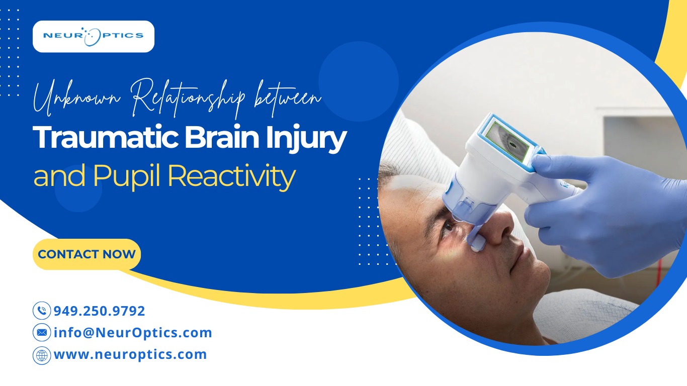
In clinical settings, pupillometers are specialized tools that measure the movement of the eyes and pupil reactivity. Therefore, the pupillary response is the movement of the eyeballs measured by the pupillometer. The electrooculogram (EOG) is a device that monitors electrical potentials on the eye’s surface. The current passing through the nerves and surrounding tissues is measured rather than the electrical activity captured by this instrument.